How is a colonoscopy done?
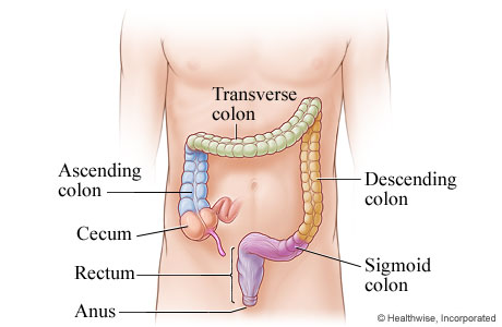
Anatomy of the colon

During a colonoscopy, the doctor will be able to look at the inside of your entire large intestine (your colon). This includes the cecum (which is attached to the small intestine and is the beginning of the large intestine), the ascending colon, the transverse colon, the descending colon, the sigmoid colon, and the rectum.
The colonoscope is placed in the colon

You will be given medicine through a needle in your vein. This is called an intravenous (IV) line. The medicine will make you sleepy. You may lie on your left side with your knees pulled up to your belly. The doctor will gently put a gloved finger into your anus. Then he or she will put the thin, flexible colonoscope in your anus and move it slowly through your colon.
The doctor looks inside the colon: Part 1

The doctor can look at the inside lining of your colon through the scope or on a computer screen hooked up to the scope.
The doctor looks inside the colon: Part 2

The doctor will look at the whole length of your colon as the scope is gently moved in and then out of your colon.
Views of a normal colon and a colon polyp

A polyp is a small growth of excess tissue that often grows on a stem or stalk. Colon polyps are growths in the colon or rectum.
Some polyps are attached to the wall of the colon or rectum by a stalk or stem (pedunculated). Some have a broad base with little or no stalk (sessile).
Current as of: May 1, 2025
Author: Ignite Healthwise, LLC Staff
Clinical Review Board
All Ignite Healthwise, LLC education is reviewed by a team that includes physicians, nurses, advanced practitioners, registered dieticians, and other healthcare professionals.